Warning: The magic method Bridge\Shortcodes\Lib\ShortcodeLoader::__wakeup() must have public visibility in /htdocs/wp-content/plugins/bridge-core/modules/shortcodes/lib/shortcode-loader.php on line 27
Information Archives - Integracell

Information

Glucose is the major energy source for mammalian cells as well as an important substrate for protein and lipid synthesis. It enters from extracellular fluid into the cell through two distinct families of structurally related glucose transporters. Once in the cells, glucose is transformed through the Glycolysis generic pathway.

The first step of glucose conversion concerns its immediate phosphorylation into glucose 6-phosphate (G6P)  through the hexokinases family : hexokinase 1,2,3 (HK1-3) and glucokinase (hexokinase 4, HK4). Primary reason for immediate glucose phosphorylation is to prevent its diffusion out of the cell. Phosphorylation that adds a charged phosphate group has two main consequences: (i) G6P because of its negative charges cannot easily diffuse through cell membrane, and (ii) the addition of the phosphoryl group begins to destabilize glucose, thus facilitating its further metabolism. The next step converts G6P to fructose 6-phosphate (F6P) by glucose phosphate isomerase (GPI). Then phosphofructokinases (PFKs, displaying three main isoforms: muscle (PFKM), platelet (PFKP), liver (PFKL)) attach the second phosphate group to F6P generating fructose 1,6 bisphosphate (F1,6BP). Aldolases (ALDO) are ubiquitous enzymes that catalyze the reversible aldol cleavage of F1,6BP to either dihydroxyacetone phosphate (DHAP) or glyceraldehyde 3-phosphate (GAP). These products consist of three-carbon units rather than six-carbon units. In humans, several aldolases isozymes, aldolases A,B,C, with different tissue distributions and kinetics exist (ALDOA in muscle and red blood cell, ALDOB in liver, kidney, and small intestine and ALDOC in brain and neuronal tissues). GAP is on the direct pathway of glycolysis, whereas DHAP is not. These compounds are isomers that can be readily interconverted : DHAP is a ketose, whereas GAP is an aldose. The isomerization of these three-carbon phosphorylated sugars is catalyzed by triosephosphate isomerase (TPI). This reaction is rapid and reversible and at equilibrium, DHAP represents 96% of the triose phosphate pool. However, the reaction proceeds readily from DHAP to GAP because the subsequent reactions of glycolysis remove this product. 

GAP is metabolized to 1,3-bisphospho-D-glycerate (1.3BPG) by glyceraldehyde-3-phosphate dehydrogenases (GAPDH1,2) enzymes. Phosphoglycerate kinases 1 et 2 (PGK1,2) catalyze the reversible transfer of a phosphoryl group from 1.3BPG to ADP which results in formation of 3-Phospho-glyceric acid (3PG). 3PG is enzymatically converted into 2-Phospho-glyceric acid (2PG) by phosphoglycerate mutase (PGAM) that displays several isoforms (PGAM1 in brain, PGAM2,3 in muscle and a multifunctional enzyme 2,3-bisphosphoglycerate mutase (BPGM) that converts 1.3BPG into 2,3-bisphospho-glycerate (2.3BPG), one of the major regulator of hemoglobin affinity for oxygen. After water release, catalyzed by different enolases (ubiquitarious α-ENO=ENO1, muscle β-ENO=ENO3, neuronal ϒ-ENO=ENO2), phosphoenolpyruvate (PEP) is formed. Then PEP is converted to pyruvic acid, the end product of glycolysis, by pyruvate kinase (PKLR in liver and RBC and PKM in muscle).

Pyruvic acid can then be either transformed into lactate through enzymatic action of lactic acid dehydrogenases (LDH, LDHA in muscle, LDHB in heart, LDHC in germ cells) or into acetylcoA by pyruvate dehydrogenase (PDH) that can be the major substrate of the tricarboxylic acid cycle in mitochondria.

Glutathione, a non-protein thiol containing molecule, is a fundamental antioxidant in plants, animals, fungi, and some bacteria and archaea, preventing damage to important cellular components caused by reactive oxygen species such as free radicals and peroxides. In mammals, the liver is the largest glutathione reservoir.

Glutathione can be found in the cell in two forms. Reduced glutathione (GSH) is a linear tripeptide of L-glutamine, L-cysteine, and glycine. As electrons are lost, the molecule becomes oxidized, and two such molecules become linked (dimerized) by a disulfide bridge to form glutathione disulfide or oxidized glutathione (GSSG). This linkage is reversible upon re-reduction. Hence, GSH is under a tight homeostatic control at both intracellular and extracellular levels. A dynamic balance is maintained between GSH synthesis, recycling from GSSG/oxidized glutathione, and utilization.

GSH synthesis involves two closely linked, enzymatically-controlled, reactions that utilize ATP. First, L-cysteine and L-glutamate are combined by gamma-glutamyl cysteinyl synthetase (GCS). Second, GSH synthetase (GSHS/GCLC) combines gamma-glutamylcysteine with glycine to generate GSH.

GSH recycling is mainly catalyzed by glutathione disulfide reductase (GSHR), which uses reducing equivalents from NADPH to reconvert GSSG to 2GSH. Thus, NADPH is mandatory to GSH recycling and is given by glucose-6-phospahte dehydrogenase (G6PD), an enzyme involved in the pentose monophosphate pathway.The reducing power of ascorbate helps to conserve systemic GSH. GSH can also resulted from conjugation of L-Cysteinyl-glycine with L-Glutamic acid, a reaction catalyzed by gamma-glutamyltranspeptidases (GGT).


Oxidative stress is well known to be involved in the pathogenesis of lifestyle-related diseases, including aging, atherosclerosis, hypertension, diabetes mellitus, ischemic diseases, neurodegeneration and/or malignancies.

Oxidative stress has long been defined as highly detrimental in humans as oxygen free radicals attack then destroy biological molecules such as lipids, proteins, and DNA. However, oxidative stress also has been shown to play a useful role in physiologic adaptation and in intracellular signal transduction regulation. Therefore, a more useful definition of oxidative stress may be “a state where oxidative forces exceed the antioxidant systems due to loss of the balance between them.” The balance is not perfect, however, so that some free radicals-mediated damage occurs continuously.

In normal physiological situations, Radical Oxygen Species (ROS) are constantly produced in our organism, where they even play several physiological roles, and their production is regulated by an efficient antioxidant defense (vitamins, oligoelements, proteins and enzymes) to prevent excessive cell damage.

All situations either physiological (prolonged exposure to pollution or sunlight, heavy consumption of alcohol and/or drugs, unbalanced physical activities, smoking, oxidant deficient diets…) or pathological  that will induce a deterioration in our antioxidant defense system will drive an overproduction of ROS in cells. The unstable nature of the latter makes them particularly reactive and capable of inflicting major cell damage by causing breaks and mutation in DNA, by inactivating proteins and enzymes, by oxidizing sugars, and by inducing lipid peroxidation among the polyunsaturated fatty acids of lipoproteins or of the plasma cell membrane. As a resultant response, altered sugars, lipids, proteins, even DNA will induce apoptosis in otherwise healthy cells or activate various genes coding for pro-inflammatory cytokines or adhesion proteins.

Pro-oxidative markers


Toxic effects of free radicals can affect either changes in intracellular redox potential, gene activation and oxidative modification of lipids, proteins and DNA. Most of the prooxidative markers belong to the latter mechanism.

Lipids are reported as one of the primary targets of ROS. Hydroperoxides have toxic effects on cells both directly and through degradation to highly toxic hydroxyl radicals. They may also react with transition metals like iron or magnesium to form stable aldehydes, such as malondialdehyde (MDA), that damage cell membranes or lipoproteins (oxidized LDLs). Secondary products of lipid peroxidation are produced among which figure either isoprostanes (prostaglandin (PG)-like substances that are produced in vivo independently of cyclooxygenase (COX) enzymes, primarily by ROS peroxidation of arachidonic acid) or another major toxic aldehyde 4-hydroxy-2,3-nonenal (HNE), which is produced by the oxidation of ω-6 polyunsaturated fatty acids (arachidonic, linolic and linolenic acids)).

Protein peroxidation can be processed either by MDA and HNE secondary oxidation of protein structures but also by metal oxidation as the reduced metal form (Fe2+, Cu2+) may reduce peroxide to hydroxyl radical or pheryl radical (Fenton’s reaction) that will attack aminoacids. Therefore metal status evaluation is important (total iron, ferritin) as well as oxidatively modified proteins. Nitrotyrosyl residua indicate the presence of increased synthesis of peroxynitrite, i.e. nitric oxide and superoxide.

Oxidative damages of DNA that are most frequently manifested by base loss and formation of abasic sites (AP site), unilateral cleavage of DNA chain and sugar modification. These modifications can also lead to DNA double strand bilateral breakage that is a highly mutagenic event. The most commonly used marker of oxidatively modified DNA molecule is 8-hydroxy-2’-deoxyguanosine (8-OHdG).

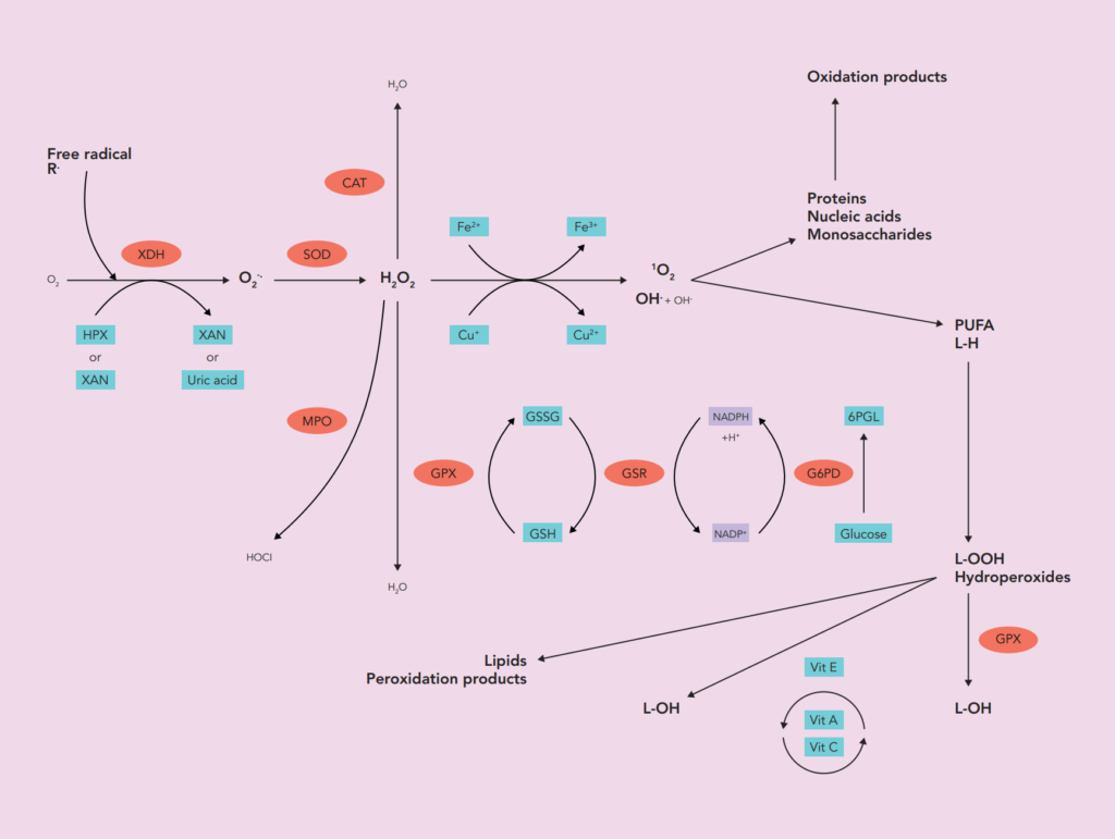
As a marker of production, activity of several enzymes aimed at generating free radicals can also be measured (xanthine oxidase (XO), myeloperoxidase (MPO), NADPH oxidase (NOX)).

Antioxidant markers


Antioxidants constitute the major defense system of our organism against ROS and free radicals. Their level can be largely modulated by nutritional diet. Because all these antioxidants do act synergically, it is important not only to measure their absolute concentration but also the ratios existing between them.

  • To evaluate antioxidant defenses level, different subset of markers must be evaluated :
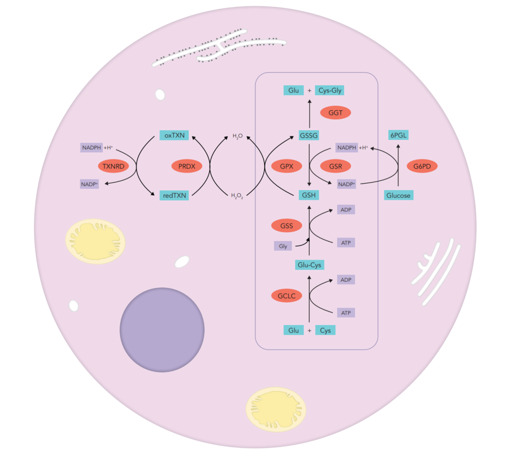
    enzymatic activity assays concerning enzymes strongly involved in free radicals and peroxide detoxification such as superoxide dismutase (SOD1, SOD2), glutathione peroxidase (GPx), catalase, thioredoxin reductase (TRx) and glutathione pathway enzymes (glucose-6-phosphate dehydrogenase (G6PD), glutathione synthase (GS) and glutathione reductase (GR))
 
  • small molecules able to scavenge free radicals such as carotenoids (vitamin A and β-carotene), vitamin C, vitamin E (α and γ), glutathione (GSH) and its oxidised product (GSSG), uric acid, bilirubin, coenzyme Q10, trace elements such as selenium that plays an important role as part of antioxidant selenoprotein